〔Research in Theory and Basic Sciences理論與基礎研究〕
In-depth observations on eye
floaters
– a challenge to ophthalmology
By Floco
Tausin
[
ABSTRACT ] For centuries, scholars try to find
an explanation for the mobile, scattered and transparent spheres and strings in
our visual field. Early on in ophthalmological tradition, the origin was
thought to be in the eye. The phenomenon was considered a disorder or
degeneration somewhere between cornea and retina. Today, eye floaters are
believed to be an opacity of the vitreous. However, careful observation of
floaters reveals properties that challenge this dominant view and call for a reconsideration
of the ophthalmological explanation.
In ophthalmology, “eye floaters” is a collective term for
vitreous opacities which are attributed to different causes. In most cases,
however, the phenomenon is considered a non-pathological (idiopathic) age-related
clouding of the vitreous. In this article, my statements on floaters refer to
this idiopathic type. According to ophthalmologists, this wide-spread symptom
occurs due to the liquefaction (synchysis)
and collapse of the collagen-hyaluronic structure of the vitreous (syneresis), which at some stage causes the
detachment of the vitreous from the retina (posterior vitreous detachment) (Sendrowski
2010). In daylight, degenerated vitreous structures which are clumped together cast
shadows on the retina and become visible in the field of vision. Supposedly,
this is what we see when we are looking at our mobile, scattered and transparent
dots and strings.

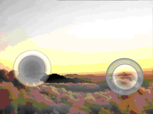
Floaters as vitreous opacities. Source:
flickr, http://www.flickr.com/photos/andrewcoulterenright/4106224/
This
ophthalmological description is the latest offshoot in a tradition recorded since
the time of Hippocrates. Over the centuries, the terms muscae volitantes (Latin, “flying flies”) or myodesopsia (Greek, “seeing fly-like corpuscles”) were used in
Greek, Arab and Western European ophthalmology to describe subjective visual
phenomena that look similar to flying flies. From the beginning, a number of
eye diseases and disorders were associated with flying flies, e.g. scotoma, cataract
or retinal detachment. This reflects the endeavor to localize and explain eye
floaters which, in turn, depends on the dominant philosophy: the ancient
natural philosophers and scholars stressed that floaters must be in the liquids
near the eye’s lens, which was taken as the main element of seeing. Later, the materialistic-mechanical
philosophy, on which early modern ophthalmology is based, promoted the notion
of floaters as physical objects
that move in the liquid of the vitreous near the retina, depending on the movement
of the eyes, consistency of the medium, gravity as well as laws of hydrodynamics. 19th century Czech
physiologist Jan Evangelista Purkinje explained the spheres and strings as fibrillae,
vessels or dead materials near the retina whose shadows were projected on the
retina when light enters the eyes. Most present-day eye doctors basically refer
to Purkinje’s description (Hirschberg 1889-1912; Plange 1990).
In
my view, this historically grown equating of spheres and strings and fly-like
visual disorders or cloudings is the result of a one-sided objective approach
and of disciplinary narrowness. To balance this, I’m going to provide some challenging
observations on floaters that I have collected in my many years of holistic research
(Tausin 2009a, 2010b). Since individual observation is my starting point and
base for my conclusions, I do not claim general validity, but I do encourage
the inclined reader to spend some time in close observation of his or her own floaters
– as a way to make my findings comprehensible.
Inconvenient
questions to ophthalmology
Where
does the morphological regularity of floaters come from?
Floaters
are dots and strings. The strings are filled with rows of dots or spheres that
are more or less clearly visible. The dots are circular and concentric; they
contain a core and a surround, viz. they are polar. The polarity is joined by a
dualism, for there are two types of dots: those with bright surround and dark
core, and those with dark surround and bright core. So we can speak of a
dualistic-polar principle in eye floaters. It’s hard to imagine that randomly
clumped vitreous fibrillae produce dots with such clear and repeated morphological
characteristics.
The two contrasting types of polar
floater spheres. Source: author.
Why are there different states of floaters?
On
closer observation, floater spheres and strings show different states over time:
one and the same sphere can appear as big and rather hazy or as small and
clearly outlined. The transition from one state to another is fluid and proceeds
in different time duration. For the sake of simplicity, I distinguish an
initial or relaxed state and a final or concentrated state. In general, it
seems that most floaters are initially relaxed, viz. bigger, closer and more
transparent; with increasing time of observation, they change into the
concentrated state. After completion of the concentration – a quick glance to somewhere
else may suffice –, the spheres and strings change back into the initial relaxed
state.
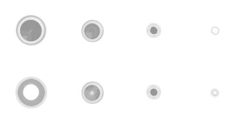
The two kinds of floater spheres in
transition from a relaxed (left) to a concentrated (right) state. Source: author.
Why do floaters start to light
up after some time of concentrated observation?
It
is interesting to realize that, in the concentrated state, the spheres and
strings increase in brilliance. Considering an energetic explanation for this,
we could say that the amount of light or energy contained in a sphere or string
does not change in the process of concentration; rather, the energy gets compressed
due to the reduction of space resulting in more brilliance (Tausin 2009a,
2010b). This effect may be influenced by “external” factors: it is encouraged
by bright lighting conditions and the distance of the focal point – the closer
the focal point, the brighter the floaters. Also, observing the spheres and
strings through the eyelashes or a pinhole in a sheet of paper lets the floaters
appear concentrated. It is important to experience, though, that the concentration
state is also reached without these aids, solely by focusing on floaters for a while;
it is quickly brought to an end by visual distraction. Thus, floaters seem to
reflect both outer and inner conditions of light and nearness, or concentration
and presence, respectively.
Ophthalmology
does not provide an explanation for the different states and the lighting up.
Eye doctors, when asked, tend to ignore the question. Some try to explain the
change in size as a result of floaters getting closer to the retina while
looking up to the sky – gravity pulls the floaters back to the retina. The
argument is unconvincing since the same effect can be observed irrespective of
whether the eyes look up to the sky or down to the ground. Others trace the
brilliance effect back to the scattering and reflectance of light. This is
supposed to happen when light strikes the floaters inside the vitreous (Tausin
2005a). The lens effect explanation implies the above-mentioned moving of
floaters inside the vitreous. It is problematic insofar as it does not take
into consideration the evident regularity of the altering of floater states
(the nearer the focal point, the brighter the floaters; the longer the observation,
the brighter the floaters). Moreover, the notion of moving dots and strings
inside the vitreous raises further questions.
Why do floaters move so
quickly if the vitreous is a jelly-like fluid?
Floaters
can be set in motion by eye movements. Doing so, they often seem to glide very
easily and with high speed across the visual field. This is all the more
surprising if we consider that the vitreous is thicker than water and described
as a gel (Ruby 2007). How can there be any particles moving so quickly and
effortlessly in a jelly-like mass? The classic answer is that the vitreous
liquefies over time and floaters become very mobile. This leads straight to the
next question.
If floaters are particles
floating in liquefied parts of the vitreous, why do we keep seeing the same
spheres and strings?
Anyone
who closely watches his or her floaters will soon become acquainted with them. For
these spheres and strings remain the same for years. Through vigorous eye movements
we may change the relative positions of the spheres and strings to one another,
but only temporarily – the floaters take their starting position soon again.
This observation contradicts the notion of free floating particles in the liquefied
parts of the vitreous – these would be whirled around with every eye movement
and take up new constellations. The medical argument goes that some floaters do
not move freely in the vitreous but are attached to the still existing vitreous
structure. From the individual observer’s perspective, there is no evidence: while
some of the strands whose ends go beyond my visual field might be attached,
other strings and all of the spheres do not seem to be attached anywhere – but
still appear in their characteristic constellations.
Why do floaters tend to sink?
Through
eye movements, we can move floaters in all directions. But as soon as we keep
our eyes still, we realize that they sink down in our visual field – the nearer
and bigger ones faster, the others more slowly. Gravity effects seem to be a
plausible explanation for this sinking of physical particles in the vitreous. The
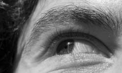
case is more complicated, though: As we know, our eyes project an upside-down
image of what we are looking at on the retina.

Source: http://www.danielng.com.au/fiwee/?p=279
(15.6.11)
If
floaters were particles close to the retina that are pulled down by gravity and
cast shadows on the retina, then I would have to see floaters rise in my visual
field. Since I do not see floaters rising but sinking, the conclusion would be
that the corresponding particles in the vitreous do not sink but rise. If that
is true, there would be other forces than gravity influencing the upward movement
of floaters.
I have
asked dozens of eye doctors about this with no convincing results. Most ignore
the fact of the inverted retinal image, or consider floaters or the retinal
image isolated from one another. Some admit that floaters have to rise in the
vitreous if we see them sinking in our visual field. This leads them to speculate
about thermodynamics (heat as lifting force) or density (floaters are lighter
than the vitreous liquid) as responsible mechanisms for that observation
(Tausin 2010a).
Further inconsistencies in ophthalmology
Why can’t eye doctors see floaters in the eyes?
If the so-called “idiopathic
eye floaters” are really clouding particles in the vitreous, then one would
think that eye doctors see them when looking in the patients’ eyes. In reality,
there is often a discrepancy between the patient’s observation of eye floaters
and the doctor’s findings in the eye. In many cases, doctors can’t see anything
while patients very clearly perceive, describe and draw their eye floaters (cf.
Weber-Varszegi et al. 2008; Tausin 2008). Then the diagnosis goes somewhat like
“age-related harmless eye floaters”, together with the advice to just ignore
them. Explanations for this discrepancy are easily found: the opacities are too
small to be relevant; the technology used is not accurate enough; the doctors
do a poor examining job; the patient is exaggerating or has a mental problem. While
there might be some truth in all these points, we also should keep in mind the
possibility that floaters are not what today’s ophthalmology claims.
It is no surprise that explanatory
innovation comes from laser surgeons. In order to treat floaters with the
Nd-YAG laser, surgeons have to localize and recognize the different floater
types very carefully. The eye doctors James Johnson and Scott Geller explain on
their websites that some floaters, especially those in young people, cannot be
seen and treated with laser. The description of these “ill-defined” floaters fits
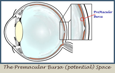
the idiopathic ones at issue. The surgeons hold the opinion that this type is not
located in the vitreous, but must be between vitreous and retina, a space
called “bursa premacularis” (Geller, n/a; Johnson n/a; cf. Tausin 2009b). This
space is potential insofar as it exists only if fluids separate vitreous from
retina. In these fluids, rests of cells or fibrillae could remain that become
visible as floaters. While the theory is not acknowledged among eye doctors –
as laser surgery of floaters is itself treated with reservation by many –, it
does not contradict the main strategy to remove floaters: vitrectomy.
Does vitrectomy prove the vitreous opacity theory of
floaters?
The
most powerful argument for the notion of floaters as vitreous opacities are the
different forms of vitrectomy, a surgery to remove and replace the whole or
parts of the vitreous. Laser surgeons assume that even bursa floaters might be removed by
vitrectomy if the vitreous is previously detached from the retina (cf. Tausin
2009b). In literature, there are cases of successful floaters-only vitrectomies
(FOV), or “floaterectomies”, in patients with “idiopathic” or “persistent”
floaters which had no or little objective correspondence (Roth et al. 2005; cf.
Tausin 2005b). In clinical studies that evaluate the outcome of vitrectomies
for floaters performed to relieve the patient’s subjective strain, patients’
satisfaction is strikingly high – around 90% (Schulz-Key et al. 2011;
Weber-Varszegi et al. 2008). This figure must not be taken as a proof for the
harmless floaters being vitreous opacities, though, for several reasons: in
these studies, it is never entirely clear what kind of floaters these patients
really saw; even if they are called “idiopathic”, patients might not have seen
the floater type at issue. Moreover, the patients’ satisfaction is influenced
by a number of factors such as visual improvement due to removing cataract and
even subjective expectancy – the latter, together with the incomprehensible
patients’ strain as a motivation to get rid of floaters, tends to turn floaterectomy
into a kind of psychotherapy (Tan et al. 2011; Tausin 2008). Also, there are
many reports of patients that have experienced floaters after vitrectomy (Schulz-Key
2011; Degenerative Vitreous Community n/a). They are explained as remaining
vitreous fibrillae or newly developed floaters. Finally, if idiopathic floaters
are no longer seen after FOV, there still might be other explanations for this.
It is conceivable that the light is channeled through the eye in a different (unstructured)
way and, therefore, stimulates the retinal neurons differently, resulting in a
vision with less or no floaters. Therefore, I suggest that the origin of floating
spheres and strings should be looked for in the activity of visual neurology
(Tausin 2009c, translation forthcoming).
Conclusion
Present-day
ophthalmology provides a frame to understand and describe the subjective visual
spheres and strings known as harmless or idiopathic eye floaters. It is a
historically grown melting pot in which floaters got associated with a number
of eye disorders. A close observation of floaters reveals properties for which the
disorder theory fails to provide a convincing explanation. Moreover,
inconsistencies within this explanatory frame itself tell us to remain critical.
The spheres and strings
are a subjective phenomenon. To study them means to be aware of that fact and
to start from individual observation. We also have to keep in mind that
perception is shaped not only by sensory data but also by our consciousness
state, mental dispositions, motivations, cultural and social environments, etc.
For example, it is my experience that size, luminosity and movement of floater
spheres and strings alter according to different consciousness states. I think that
the understanding of experiences like this are crucial in the search of a more
reasonable understanding of floaters (Tausin 2009a). The subjective approach does
not replace but complement and inform physiological research. For example, the
observations presented in this article suggest to consider the role of the
visual nervous system in the process of seeing floaters.
References:
The pictures are taken from image hosting websites,
from scientific publications (online and print) and/or from my own collection
(FT). Either they are licensed under a Creative Commons license, or their copyright
is expired, or they are used according to the copyright law doctrine of ‘Zitatrecht’,
‘fair dealing’ or ‘fair use’.
n/a (n/a): Floaters only
vitrectomy. In: Degenerative Vitreous Community. http://floatertalk.yuku.com/forums/2/Floaters-Only-Vitrectomy#.Tfh-e0djmy4
(15.6.11)
Geller,
Scott (n/a):
Hirschberg,
Julius (1899-1918): Geschichte der Augenheilkunde. In: Handbuch der gesamten
Augenheilkunde, ed. by
Johnson,
James H. (n/a): Vitreous Floater Solutions. vitreousfloatersolutions.com
(12.11.09)
Plange,
Hubertus: Muscae
volitantes – von frühen Beobachtungen zu Purkinjes Erklärung, in: Gesnerus 47, 1990,
S. 31-44
Roth, M. et al. (2005): Pars-plana-Vitrektomie
bei idiopathischen Glaskörpertrübungen, in: Klinische Monatsblätter der
Augenheilkunde 222: 728-732
Sendrowski, David P.;
Bronstein, Mark A. (2010): Current treatment for vitreous floaters. In:
Optometry 81: 157-161
Schulz-Key, Steffen et
al. (2011): Longterm follow-up of pars plana vitrectomy for vitreous floaters:
complications, outcomes and patient satisfaction. In: Acta Ophthalmologica 89:
159-165.
Tan, H. Stevie et al. (2011):
Safety of vitrectomy for floaters. In: American Journal of Ophthalmology. 151,
no. 6: 995-98.
Tausin, Floco (2010a):
Aus der Wissenschaft. Von aufsteigenden und absteigenden Mücken. In: Ganzheitlich
Sehen 1. http://www.mouches-volantes.com/news/newsfebruar2010.html
(9.6.11)
Tausin, Floco (2010b).
Eye Floaters. Floating spheres and strings in a seer’s view. In: Holistic
Vision 2. http://www.eye-floaters.info/news/news-june2010.html
(15.12.10)
Tausin, Floco. (2009a):
Mouches Volantes. Eye Floaters as Shining Structure of Consciousness.
Tausin, Floco (2009b):
Aus der Wissenschaft: Mouches volantes nicht im Glaskörper? In: Ganzheitlich
Sehen 4 (Dezember). http://www.mouches-volantes.com/news/newsdezember2009.html
(11.6.11)
Tausin, Floco (2009c):
Mouches volantes – Glaskörpertrübung oder Nervensystem? In: ExtremNews. http://www.extremnews.com/berichte/gesundheit/e01c12cc1d3c89f
(22.12.09)
Tausin, Floco (2008): Neues aus der Wissenschaft: “Floaterektomie” als
Psychotherapie? In: Ganzheitlich Sehen 3 (Oktober). http://www.mouches-volantes.com/news/newsoktober2008.html
(15.6.11)
Tausin, Floco (2005a): Neues aus der Augenheilkunde: Nicht
repräsentative Umfrage unter Augenärzten zum Thema “Mouches volantes”. In:
Ganzheitlich Sehen. http://www.mouches-volantes.com/news/newsaugust2005.html
(9.6.11)
Tausin, Floco (2005b). Neues aus der Augenheilkunde: Klinische Studie
über die Pars-plana-Vitrektomie bei Glaskörpertrübungen. In: Ganzheitlich
Sehen. http://www.mouches-volantes.com/news/newsnovember2005.html
(15.6.11)
Weber-Varszegi, V. et
al. (2008): „Floaterektomie“ – Pars-Plana-Vitrektomie wegen Glaskörpertrübungen,
in: Klinisches Monatsblatt Augenheilkunde 225: 366-369
The author:
The name Floco Tausin is a pseudonym. The
author is a graduate of the Faculty of the Humanities at the
Contact:
floco.tausin@eye-floaters.info
The book:
Mouches Volantes. Eye
Floaters as Shining Structure of Consciousness‘.
(Spiritual Fiction. ISBN: 978-3033003378.
Paperback, 15.2 x 22.9 cm / 6 x 9 inches, 368 pages).
Floco Tausin tells the
story about his time of learning with spiritual teacher and seer Nestor, taking
place in the hilly region of
«Mouches Volantes»
explores the topic of eye floaters in a much wider sense than the usual medical
explanations. It merges scientific research, esoteric philosophy and practical
consciousness development, and observes the spiritual meaning and everyday life
implications of these dots and strands.
«Mouches Volantes» – a
mystical story about the closest thing in the world.
洽詢地址Add.: 2712
San Gabriel Boulevard
Rosemead, CA 91770 U.S.A.
電話Tel.: (626)
288-1199
傳真Fax: (626)
288-4199